Optical Diagnosis
Using optics to discover the mysteries of the body below are examples of the power of microscopic techniques in bio-medicine.
Looking Inside Your Eye
Here, a three-dimensional recording of a human lens is made using a microscope called a Scheimpflug Camera to identify imperfections, or cataracts, in the human eye. The lighter colors appearing in the video are the more opaque, or imperfect, sections of the lens. This is an example of the merging of optical and computer technologies to measure the scattering of light inside the eye effectively mapping the inside of the living eye. Click on the image to view a video (1.9MB) in a Quicktime format.
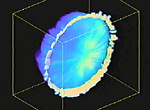
The eye lens is from a living patient.
Contributor: Barry Masters
Source material: Optics Express, Volume 3, Issue 9, 332-338 October 1998
Note: The colors seen in these videos are psuedocolors - colors assigned to represent different depths, opacities and layers in the tissue. They are not the actual colors in the living tissue.
Here, a three-dimensional recording of a section of a rabbit cornea is made using reflected light confocal microscopy. Two cell layers and a corneal nerve (orange-red) are recorded (not to be confused with the optic nerve which connects nerve cells in the retina with the brain). Click on the image to view a video (2.0MB) in a Quicktime format.
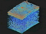
The rabbit cornea is from a living, excised rabbit eye.
Contributor: Barry Masters
Source Material: Optics Express, Volume 3, Issue 9, 351-355 October 1998
Note: The colors seen in these videos are psuedocolors - colors assigned to represent different depths, opacities and layers in the tissue. They are not the actual colors in the living tissue.
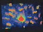
Here, a close-up section of the corneal nerve and surrounding cell nuclei is recorded. Click on the image to view a video (1.2MB) in a Quicktime format.
Contributor: Barry Masters
Source Material: Optics Express, Volume 3, Issue 9, 351-355 October 1998
Note: The colors seen in these videos are psuedocolors - colors assigned to represent different depths, opacities and layers in the tissue. They are not the actual colors in the living tissue.
Looking Behind Your Eye
Optics, and in this case lasers, are used to record the area behind the living eye. Here, a scanning laser confocal microscope is used to visualize the human fundus (the back portion of the eyeball) from near the retina surface to deep within the optic nerve. Click on the image to view a video (2.5MB) in a Quicktime format.
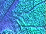
The optic nerve is from a living patient.
Contributor: Barry Masters
Source Material: Optics Express, Volume 3, Issue 10, 356-359 November 1998
Note: The colors seen in these videos are psuedocolors - colors assigned to represent different depths, opacities and layers in the tissue. They are not the actual colors in the living tissue.
Looking Inside Your Skin
Here optics is used to record the different layers of skin on a personís forearm using two different techniques.Here is a graphic showing a cross-section of human of skin.
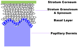
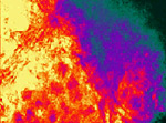
Recording of the Same Section of Skin Using Different Techniques
The first technique uses confocal reflected light microscopy to measure thin "optical sections" of skin. In other words, the reflection of light from the layers of skin are being measured. Click on the image to view a video (700kb) in a Quicktime format.
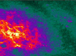
Here, the same section of skin from the forearm is being measured with multiphoton excitation microscopy to measure the "optical sections" of skin. However, in contrast, the fluorescence of coenzymes are being recorded and not the reflections from the layers of skin. Notice the greater detail and depth recorded using this method of measurement on the same section of skin. Click on the image to view a video (528kb) in a Quicktime format.
The skin measured is that of the author Dr. Barry Masters.
Contributors: Barry Masters, Peter So
Source Material: Optics Express, Volume 8, Issue 1 2-10 January 2001
Note: The colors seen in these videos are psuedocolors - colors assigned to represent different depths, opacities and layers in the tissue. They are not the actual colors in the living tissue.
Using Light to Look at the Brains
If you shine a flashlight onto your hand in a dark room, you can clearly see that light can travel inside your hand and be transmitted through your hand. This is why light can be used to "look" at the brain inside the head. The light that makes it back to the skin to be detected after traveling through the skull and illuminating the brain carries important information about the blood flow and the oxygenation of the brain.
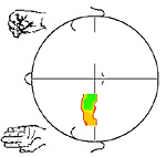
Mapping of blood flow in the brain.
By illuminating the head using low-power laser diodes and collecting the reflected light with sensitive optical detectors, we can identify regions of the brain that are activated, for instance because the subject is moving a hand. In particular, a right hand squeeze will cause an increase of blood flow on the left motor cortex, while a left hand squeeze will increase the blood flow in the right motor cortex. The brain area that responds to hand squeezing is on the opposite (or contra lateral) side of the squeezed hand.
In the video you can see that the blood flow (measured with the optical signals collected on the head) starts to increase in the contra lateral brain cortex a few seconds after the hand has started moving. When the hand stops moving, the blood flow goes back to normal.
The colors in the movie do not have a particular meaning. They just indicate the level of increase of the blood flow (red means small increase, blue means large increase). This representation is called "false-color imaging." Click on the image to view a video (400kb) in a Quicktime format.


